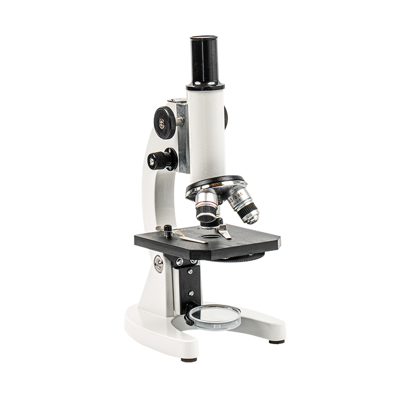
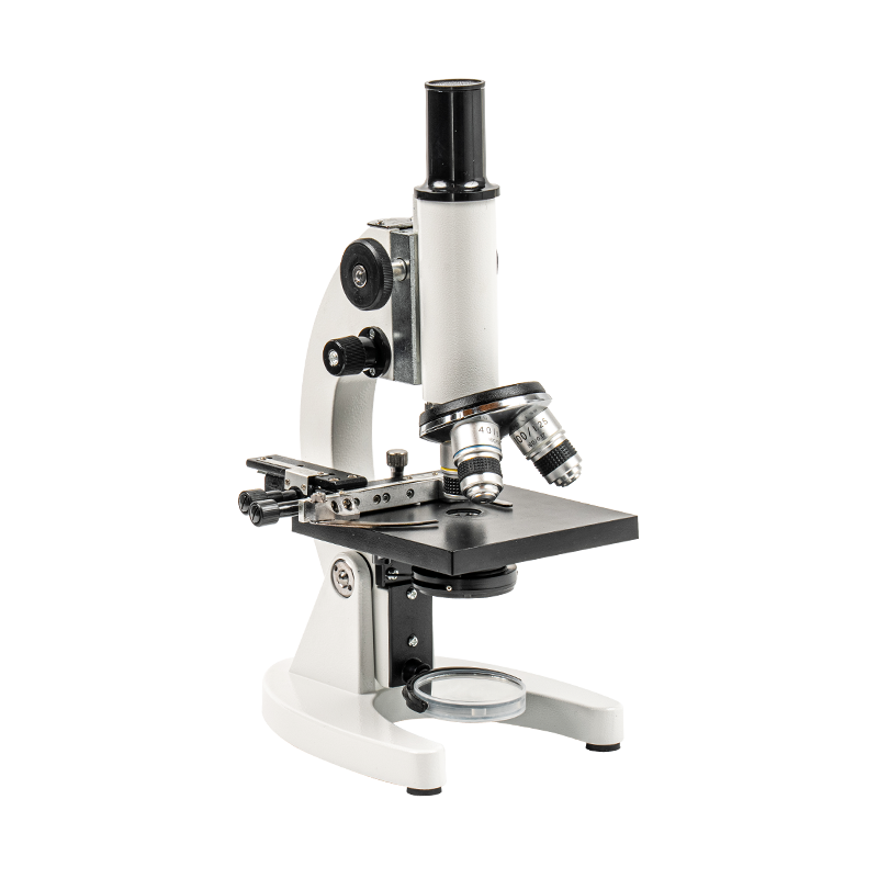
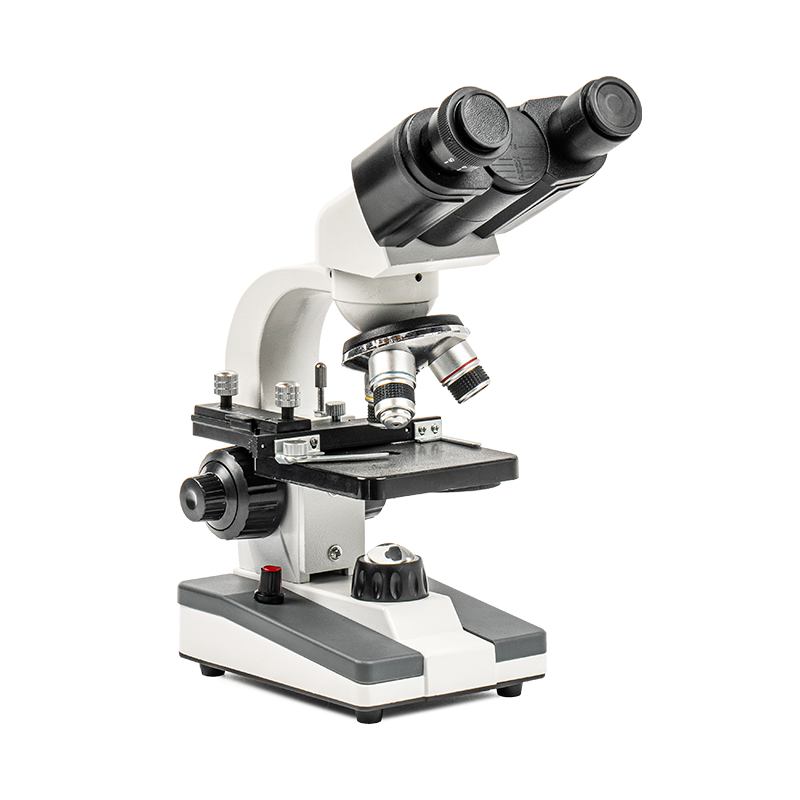
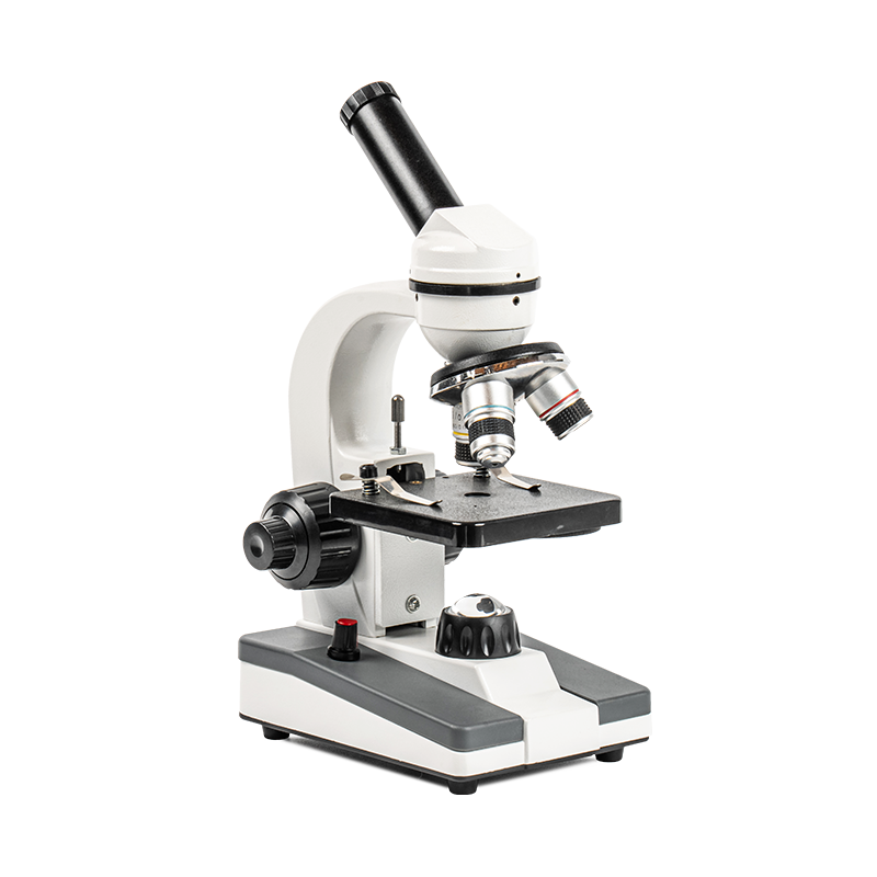
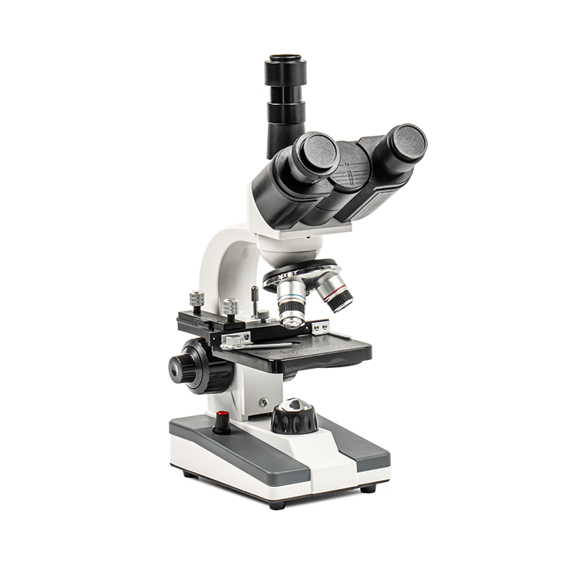
The phase contrast microscopy principle in trinocular microscope is based on the interference and phase difference of light waves, which significantly improves the contrast and clarity.
In an optical microscope, light from a light source is irradiated onto a sample through an objective lens. Different parts of the sample scatter or transmit light in different ways. When the light wave passes through the sample, a phase difference will occur in the optical path of the transparent area and the opaque area. When observing transparent samples, traditional microscopes often cannot distinguish the details of the sample well because of the lack of sufficient contrast.
Phase contrast microscopy technology enhances this phase difference by introducing a phase plate or phase ring into the light path. The phase plate has a specific thickness and refractive index that can change the phase of light waves passing through different parts of the sample. The light that passes through the phase plate is combined with the light that does not pass through the phase plate to form an interference pattern, which significantly improves the contrast of the image.
Key components
Phase plate: Phase contrast microscopes are usually equipped with phase plates, which are designed to enable coherent interference of light waves of different phases. The thickness and shape of these phase plates are precisely calculated to optimize the imaging effect under specific wavelengths of light.
Light source: A high brightness and stable light source can provide enough light to ensure a clear image of the sample during phase contrast imaging.
Objectives: Phase contrast microscopes usually use specially designed phase contrast objectives that can maximize the use of phase contrast technology during imaging. The design of phase contrast objectives takes into account the refractive index changes of the sample to ensure high quality images.

 English
 Español
 عربى
عربى 中文简体
中文简体